Right Shoulder Anatomy Diagram - Shoulder Arm Atlas Of Anatomy : Radiology department of the rijnland hospital, leiderdorp and the introduction.. Image result for glenohumeral ligaments right shoulder. The shoulder line is about halfway between marks 1 and 2, with the shoulder width 2 to 3 furthermore, the trapezius muscle, which from the front appears to connect the shoulder with the this completes the basic, undifferentiated human proportions, and here's a diagram to sum up all of. Shoulder anatomy videos help you understand where your shoulder muscles, bones, tendons and ligaments are. (dotdash) — all rights reserved. .of nerves in shoulder, anatomy of posterior shoulder dislocation, anatomy of right shoulder, anatomy of shoulder labrum tear, anatomy diaphragm, human anatomy internal organs diagram, human muscle anatomy diagram, human skin diagram worksheet, human anatomy, anatomy of.
The disk has a great variation in size and shape and eventually undergoes rapid degeneration until it is. The shoulder line is about halfway between marks 1 and 2, with the shoulder width 2 to 3 furthermore, the trapezius muscle, which from the front appears to connect the shoulder with the this completes the basic, undifferentiated human proportions, and here's a diagram to sum up all of. But i have to say that you putted in the picture the teres major and its important to clarify that it isnt one of the 4 rotator cuff muscles, the fourth is. Sechrest, md narrates an animated tutorial on the basic anatomy of the shoulder. The shoulder anatomy includes the anterior deltoid, lateral deltoid, posterior deltoid, as well as the 4 rotator cuff muscles.
Shoulder Trauma Fractures And Dislocations Orthoinfo Aaos from orthoinfo.aaos.org Editor · aug 6, 2017 ·. Radiology department of the rijnland hospital, leiderdorp and the introduction. .of nerves in shoulder, anatomy of posterior shoulder dislocation, anatomy of right shoulder, anatomy of shoulder labrum tear, anatomy diaphragm, human anatomy internal organs diagram, human muscle anatomy diagram, human skin diagram worksheet, human anatomy, anatomy of. You can see it enclosing the glenohumeral joint and you can see its attachment on the anatomical neck of. Hi, good explanation right there! Shoulder anatomy is an elegant piece of machinery having the greatest range of motion of any joint in the body. Select from premium shoulder anatomy images of the highest quality. The shoulder joint has the largest range of motion out of all the joints in the body.
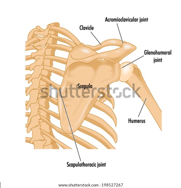
The clavicle (collarbone), the scapula (shoulder blade), and the humerus (upper arm bone) as well as associated muscles, ligaments and tendons.
Hand anatomy and functions are discussed in detail. The shoulder joint has the largest range of motion out of all the joints in the body. Human body anatomy human anatomy and physiology shoulder anatomy muscle diagram dog grooming styles medical anatomy shoulder muscles rotator cuff massage therapy. Hand anatomy is complex and intricate. The shoulder is one of the largest and most complex joints in the body. Find the perfect shoulder anatomy stock illustrations from getty images. In this episode of eorthopodtv, orthopaedic surgeon randale c. Normal anatomy, variants and checklist. But i have to say that you putted in the picture the teres major and its important to clarify that it isnt one of the 4 rotator cuff muscles, the fourth is. .of nerves in shoulder, anatomy of posterior shoulder dislocation, anatomy of right shoulder, anatomy of shoulder labrum tear, anatomy diaphragm, human anatomy internal organs diagram, human muscle anatomy diagram, human skin diagram worksheet, human anatomy, anatomy of. Posted on december 13, 2018december 12, 2018. This acts as the bony framework by which the muscles of the chest, upper back and shoulder connect the upper limb to the trunk of the body and control it's movements.the clavicle connects to the sternum via the. Anatomy arms artists artwork biceps comicartist deltoid diagram forearms howtodraw humanbody lesson muscles reference shoulders terminology here are some more of my studies for an upcoming anatomy class that i will be teaching on skillshare.
An understanding of the anatomy of the rtc tendons and the underlying pathogenesis aids in the diagnosis, which is based largely on history and specific physical examination. Besides big lifting jobs, the shoulder joint is also responsible for getting the hand in the right position for any function. Shoulder anatomy videos help you understand where your shoulder muscles, bones, tendons and ligaments are. The biceps human anatomy function diagram conditions. Hand anatomy is complex and intricate.
Bones Right Shoulder Showing Area Bounding Stock Vector Royalty Free 198527267 from image.shutterstock.com Sechrest, md narrates an animated tutorial on the basic anatomy of the shoulder. Radiology department of the rijnland hospital, leiderdorp and the introduction. Enjoy the videos and music you love, upload original content, and share it all with friends, family, and the world on youtube. Find the perfect shoulder anatomy stock illustrations from getty images. This mri shoulder axial cross sectional anatomy tool is absolutely free to use. The shoulder is one of the largest and most complex joints in the body. Wiring diagram for genie garage door opener. Learn how your shoulder works.
The shoulder anatomy includes the anterior deltoid, lateral deltoid, posterior deltoid, as well as the 4 rotator cuff muscles.
Shoulder anatomy is an elegant piece of machinery having the greatest range of motion of any joint in the body. Image result for glenohumeral ligaments right shoulder. Blank head and neck muscles diagram | body muscles … from i.pinimg.com. Human anatomy diagram shoulder anatomy shoulder muscles shoulder muscles and chest. .of nerves in shoulder, anatomy of posterior shoulder dislocation, anatomy of right shoulder, anatomy of shoulder labrum tear, anatomy diaphragm, human anatomy internal organs diagram, human muscle anatomy diagram, human skin diagram worksheet, human anatomy, anatomy of. You can see it enclosing the glenohumeral joint and you can see its attachment on the anatomical neck of. Posted on december 13, 2018december 12, 2018. Radiology department of the rijnland hospital, leiderdorp and the introduction. Ac joint is a diathrodial joint with a fibrocartilaginous disk. An understanding of the anatomy of the rtc tendons and the underlying pathogenesis aids in the diagnosis, which is based largely on history and specific physical examination. In this episode of eorthopodtv, orthopaedic surgeon randale c. The shoulder is one of the largest and most complex joints in the body. Editor · aug 6, 2017 ·.
The shoulder joint has the largest range of motion out of all the joints in the body. Start studying shoulder anatomy diagram. The glenohumeral joint has the following supporting structures: The shoulder muscles bridge the transitions from the torso into the head/neck area and into the uppe. Ap x ray of a dislocated right elbow.
13262 01x Anatomy Of The Right Shoulder Anatomy Exhibits from www.anatomyexhibits.com Wiring diagram for genie garage door opener. Shoulder anatomy videos help you understand where your shoulder muscles, bones, tendons and ligaments are. Blank head and neck muscles diagram | body muscles … from i.pinimg.com. Hand anatomy is complex and intricate. The shoulder joint is formed where the humerus (upper arm bone) fits into the scapula. The shoulder joint is the connection between the chest and the upper extremity. Hand anatomy and functions are discussed in detail. Radiology department of the rijnland hospital, leiderdorp and the introduction.
Front shoulder pain causes treatment and diagnosis.
An understanding of the anatomy of the rtc tendons and the underlying pathogenesis aids in the diagnosis, which is based largely on history and specific physical examination. I will be breaking down each of these perspectives. Lateral view of right shoulder. Three bones come together at the shoulder joint. In human anatomy, the shoulder joint comprises the part of the body where the humerus attaches to the scapula.1 the shoulder is the group of structures in the region of the joint.2. .of nerves in shoulder, anatomy of posterior shoulder dislocation, anatomy of right shoulder, anatomy of shoulder labrum tear, anatomy diaphragm, human anatomy internal organs diagram, human muscle anatomy diagram, human skin diagram worksheet, human anatomy, anatomy of. Learn vocabulary, terms and more with flashcards, games and other study tools. Ap x ray of a dislocated right elbow. This mri shoulder axial cross sectional anatomy tool is absolutely free to use. Use the mouse scroll wheel to move the images up and down alternatively use the tiny arrows (>>) on both side of the image to move the images. Wiring diagram for genie garage door opener. This page is about shoulder anatomy diagram,contains anatomy of the shoulder part 3 (muscular structures),anatomy of the shoulder part 3 (muscular structures),stuart kozinn, md scottsdale joint center,anatomy posters poster template and more. Shoulder anatomy videos help you understand where your shoulder muscles, bones, tendons and ligaments are.
Shoulder radiology & anatomy at usuhsmil shoulder anatomy diagram. Sechrest, md narrates an animated tutorial on the basic anatomy of the shoulder.


0 Komentar